Therapeutic Ultrasound

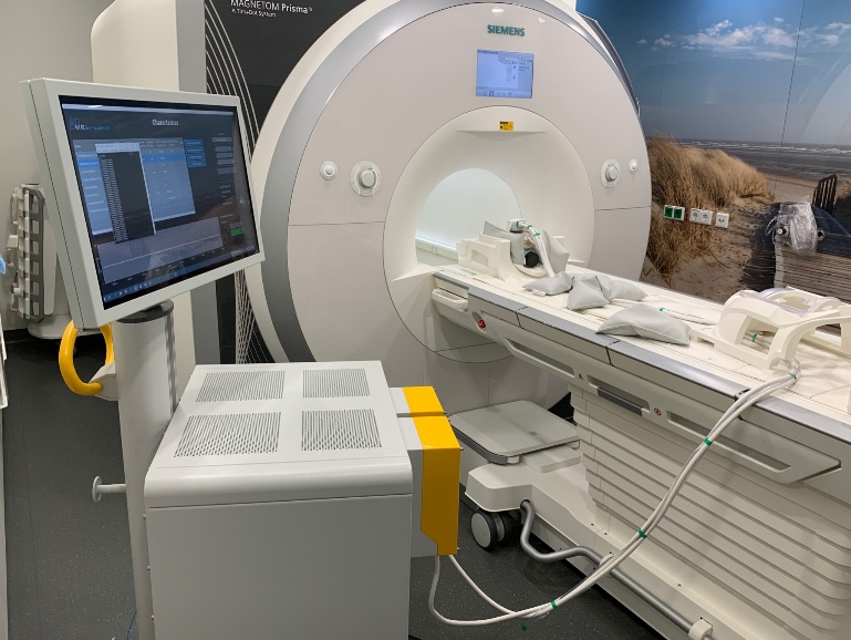
Ultrasound therapy platform for transcranial neurostimulation with volumetric beam steering

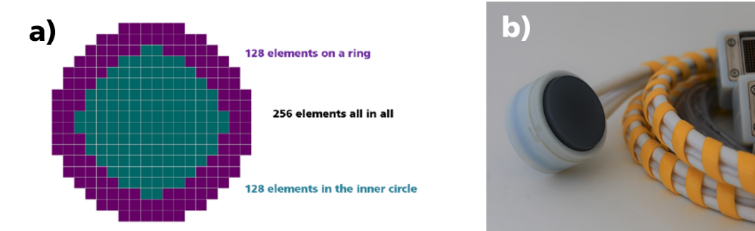
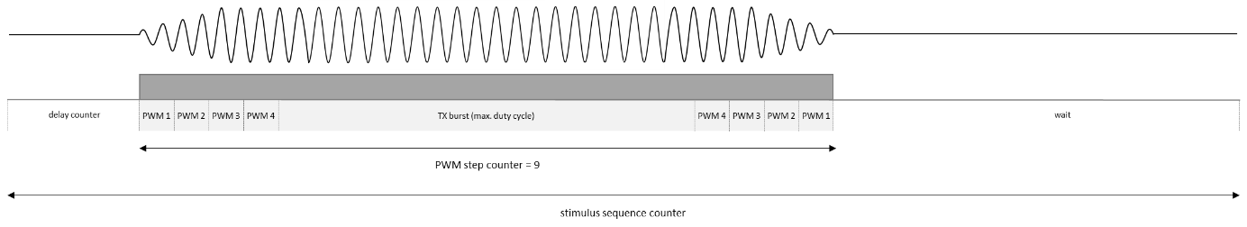
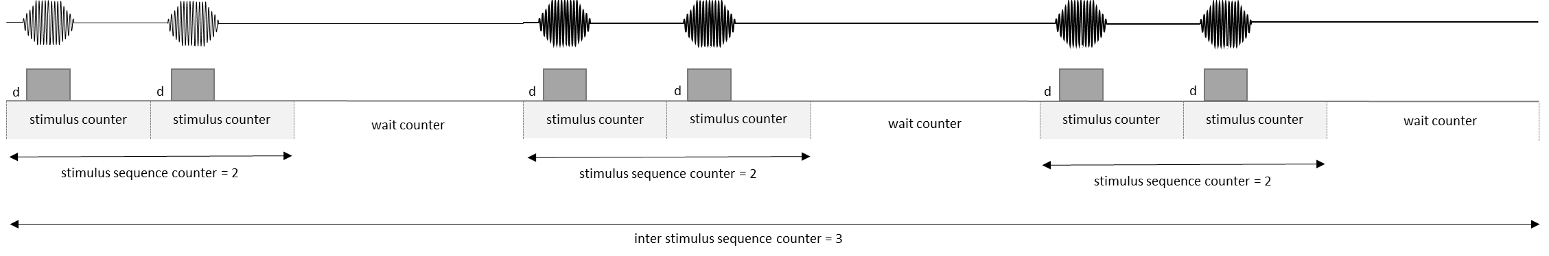
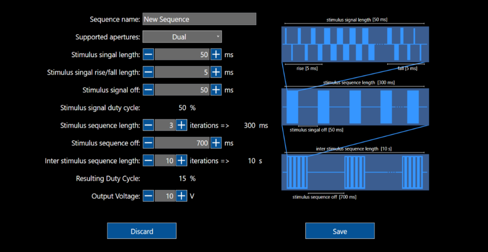
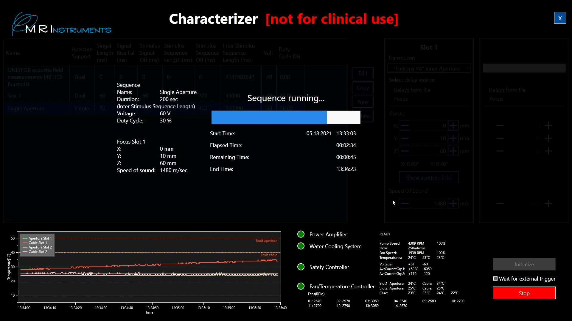
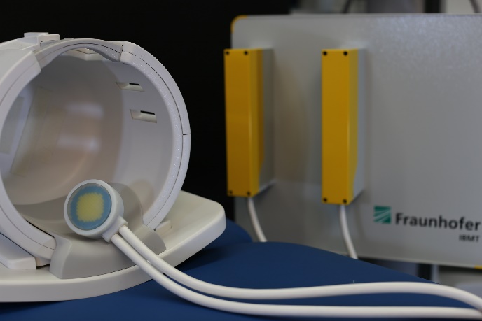
This ultrasound system for transcranial neurostimulation offers a versatile therapy solution that can function independently or in conjunction with an MR scanner for brain stimulation. It employs high-duty-cycle ultrasound signals with a specific transmission pattern directed towards a defined focal point, enabling the exploration of ultrasound sonication parameters for various target applications.
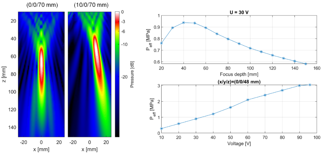
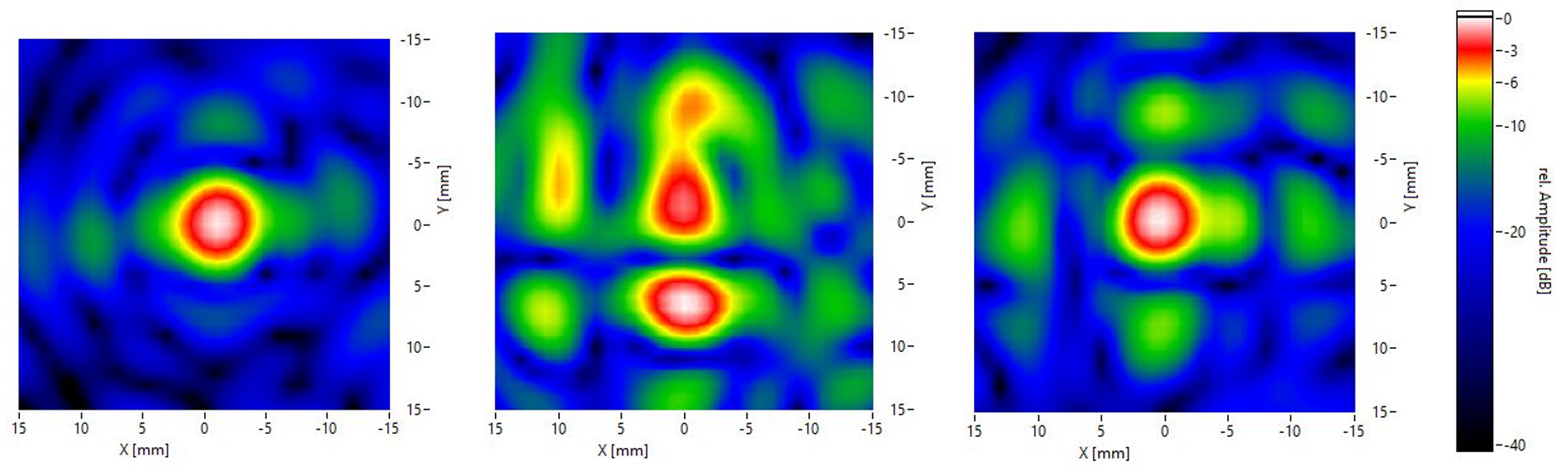
By leveraging the system's advanced 3D beam steering capabilities, it becomes possible to sonicate different brain regions, both superficial and deep. This flexibility allows for stimulating specific targets like the hypocampus, amygdala, subthalamic nucleus (STN), and motor cortex, among others. Additionally, the system acoustic power levels can be adjusted to meet the requirements of diverse applications beyond neurostimulation, including blood-brain barrier (BBB) opening.
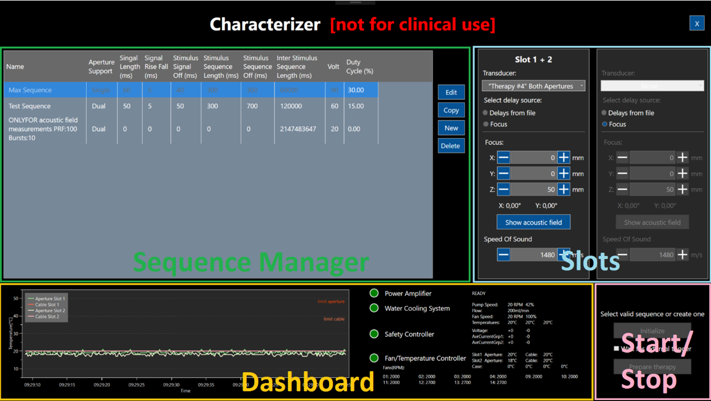
With its user-friendly graphical interfaces, the system can function as a standalone neurostimulation device. Moreover, it offers integration possibilities into custom applications through a software development kit that supports multiple programming languages.


© Fraunhofer IBMT.
Figure: (a) layout of transducer elements in sub-apertures (inner circle and outer ring, each of the apertures wired through a separate connector) (b) matrix array after integration in housing.

Targeting with volumetric transmit focus beamforming

The ultrasound system utilizes circular 2D matrix arrays consisting of 256 elements, with center frequencies of 250 kHz and 500 kHz. These arrays are arranged in a rectangular grid and divided into two separate sections: an inner circle and an outer ring. To ensure extended sonication cycles and high applied power, the transducer incorporates rear-side cooling for its active elements. The materials used are carefully selected for optimal compatibility with magnetic resonance imaging (MRI), minimizing any potential artifacts in the MRI images.

The transducer arrays are driven by a custom-designed multi-channel ultrasound transmit stage with 256 individual transmit paths. This stage generates focused, high-energy ultrasound for therapeutic applications, utilizing customizable transmission patterns. By applying unique phase differences to each transmit path, the system enables volumetric beam steering, allowing precise targeting of specific volumes of interest. This feature enhances the flexibility of transducer placement on the surface of the skull. The integrated power pulsers generate bipolar (tri-state) transmission signals with amplitudes of up to 180 Vpp (+/- 90 V) and can deliver electrical power of up to 9 W per channel for several seconds during ultrasound stimulation sequences.

The system provides two connectors, each capable of driving 128 transducer elements. Users can employ both connectors to sonicate using both the inner and outer apertures simultaneously or connect multiple individual transducers to the system. This functionality allows for cross-beam applications by driving the inner apertures of two transducers, enabling research in various configurations using the system's capabilities.


© Fraunhofer IBMT.
Figure: Ultrasound stimulation sequences with individual programming layers to support functional stimulation patterns.

Functional stimulation sequencing

The system offers versatile options for defining pulse/pause ratios at multiple levels, allowing the generation of complex sequence patterns for stimulation, particularly crucial in neurostimulation. By transmitting pulsed burst signals with consistent pulse/pause ratios over specific time intervals, the system enables the creation of intricate stimulation sequences not only at a single spot over time but also facilitates the setup of multiple focal spots that can be sequentially stimulated to expand the focal area or target multiple regions simultaneously. This flexibility enhances the system's ability to tailor stimulation according to specific requirements and applications.


© Fraunhofer IBMT.

Generated acoustic intensities

The system can generate high acoustic intensities, with a focal pressure (peak-to-peak) up to 8 MPa when focusing on a specific point, such as (0 mm / 0 mm / 48 mm), resulting in a focus size of approximately 3 mm x 20 mm (lateral / axial dimensions).
The pressure and focus size are influenced by the focus position, with the focus size increasing as the z-position is adjusted. The array allows for steering within a range of approximately +/- 20°, providing flexibility in directing the ultrasound beam.
To ensure adherence to clinically relevant limits, the system incorporates safety measures. It can restrict the output voltage and/or the duty cycle of stimulus sequences based on guidelines and limits set by regulatory bodies such as the FDA, ITRUSST, and additional applicable standards.


System setup & MR-optimization

The therapy system is seamlessly integrated into a cart, allowing for its convenient use in non-MR environments while connected to a PC or laptop. Every component of the system is meticulously designed and constructed to ensure MR optimization, aiming to minimize any additional interference with the MR scanner when operated in close proximity. It offers both triggered (sequential) and parallel operation modes, providing flexibility in its usage alongside the MR scanner.
When the system is employed in combination with an MR scanner, an antimagnetic cart is available, which can be controlled using an optional MR-optimized touchscreen and a remote-control unit. This configuration allows the cart to be positioned next to the patient's bed within the MR scanner room, while the power supply and water-cooling unit are situated in a corner of the room.
To establish a physical connection between the equipment cart inside the scanner room and the remote-control unit, an optical fiber is employed, passing through a feed-through panel into the MR scanner room. This setup ensures efficient and reliable communication between the components of the system while maintaining the necessary MR compatibility.

© Fraunhofer IBMT.
Figure: Example of flexible transducer coupling pad (left), mounting adapter to attach it to a transducer (middle) and MR-visible tracker adapter (right).

Mounting and adapters

To ensure consistent and reliable acoustic coupling, gel pads of varying thicknesses are available, enabling effective coupling even on non-planar surfaces. A fixation ring is provided to securely attach the coupling pad in front of the transducer's aperture. Additionally, a head-fixation belt with a specially designed adapter for the transducer housing has been developed, facilitating stable positioning during procedures.

To accurately track the position and orientation of the transducer in relation to the MR planning data sets, MR-visible trackers are integrated into the transducer housing. This feature allows for precise alignment and coordination between the ultrasound system and the MR imaging. Furthermore, the system offers an option for the integration of optical trackers on the probe, providing additional tracking capabilities for enhanced spatial awareness and accuracy.

Safety features

Throughout the generation of acoustic output, the system maintains continuous monitoring of the temperature on the active transducer surface and the ultrasonic transducer cable. To ensure adherence to safety standards, the system incorporates a dedicated safety circuit. This circuit is designed to automatically halt sonication if the temperature surpasses predefined threshold values for either the transducer aperture or the cable prioritizing user safety during operation.

© Fraunhofer IBMT.
Figure: User interface of software system „Characterizer” with the basic areas of configurations.
© Fraunhofer IBMT.
Figure: User interface part of “Characterizer” to define transmit sequences.
© Fraunhofer IBMT.
Figure: User interface of „Characterizer” showing progress and monitoring during acoustic output.

Software systems

The software module called "Characterizer" serves as a foundation for experimental applications. It offers a user-friendly interface for defining transmit signals, including selecting a focal point, specifying temporal patterns (such as pulse/pause ratio, PRF, frequency), and applying user-defined phase offsets or delays. This software module does not provide clinical workflow guidance and does not support loading patient-specific data for therapy planning. It is primarily intended for system characterization, compatibility experiments, and standalone phantom experiments. A MR guided workflow software framework is available to be adapted to specific requirements.

Moreover, the system offers a Software Development Kit (SDK) that provides a direct programming interface, allowing integration into other software systems. The interface is available in the C++ programming language and enables basic parameter setup and device operation. It includes system health monitoring and error handling. It should be noted that this interface is considered experimental and intended for advanced research purposes.
Binary libraries are provided, and source code samples demonstrate how to integrate the system, allowing for the development of custom applications and specific partner integration. This flexibility empowers users to create tailored solutions based on their specific requirements and research needs.

© Fraunhofer IBMT.
Figure: Example of refocusing in aberration correction: Acoustic pressure distribution (X/Y-Scan at constant depth Z = 70 mm) without skull bone (left); with skull bone induced aberration and defocusing (middle) and with skull bone including aberration correction performing refocusing (right).

Skull bone aberration correction

The system incorporates a specialized feature for correcting defocusing artifacts caused by skull bone, ensuring optimal ultrasound focus. This correction is applied by utilizing the provided transducer location and extracting information about the skull bone interface beneath it. Through experimental evaluation, it has been observed that this aberration correction not only enhances the maximum acoustic intensity but, more importantly, repositions the actual focus spot to align with the intended volume of interest. The results highlight the effectiveness of the correction in improving both the intensity and accuracy of the ultrasound focus, enabling more precise targeting during procedures.

Certification and testing

The development, documentation, and testing of the system and its components adhere to the standards outlined in the Medical Device Regulation (MDR 2017/745). While the system offers the flexibility of a research device, it is important to note that it is not CE-labeled as a standalone medical device. However, we ensure compliance through the implementation of technical risk management procedures and the provision of test reports conducted by certified laboratories. These test reports cover various aspects, including electrical safety, electromagnetic compatibility, acoustic safety, and the software development process. These measures demonstrate our commitment to upholding stringent quality standards and ensuring the safety and reliability of the system.


Simulation/Advance Development


Software Development/System Integration


Electronics Engineering


Transducer Engineering


Manufacturing Technology (ISO 9001 & 13485)

General Information

about the Department of Ultrasound

Technical Equipment

of the Department of Ultrasound

Please contact us

We offer bachelor and master thesis, internships and practical semester work for students.

Contact Press / Media

Kerstin Knobe

Secretary

Fraunhofer-Institut für Biomedizinische Technik IBMT
Ensheimer Straße 48
66386 St. Ingbert

Phone +49 6897 9071-301

Fax +49 6897 9071-302