Fraunhofer Institute for Biomedical Engineering
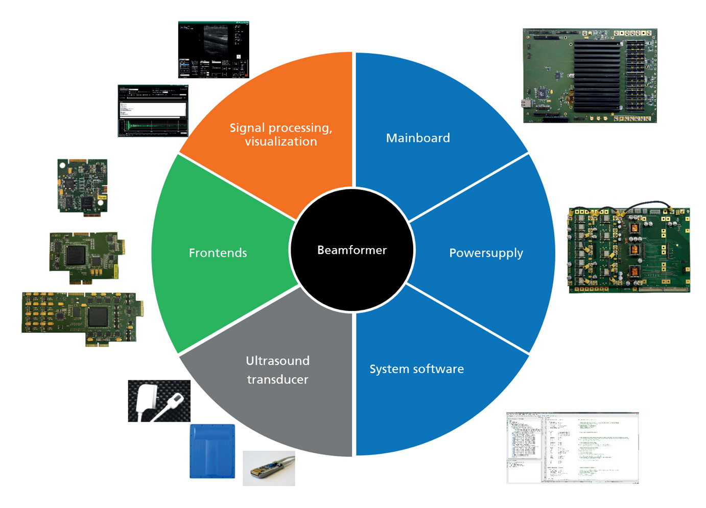
System Description

Diagnostic imaging quality of ultrasound systems is defined by the beamforming characteristics of the ultrasonic device. Modern ultrafast ultrasound plane wave imaging, dynamic focusing, steering, amplitude weighting, pulse coding and controlling the size of the aperture of an array probe are the techniques which are used to form the acoustic beam. Especially for research and development it is necessary to have complete control possibilities over the parameters that determine the geometry, the direction, the number and the acoustical properties of the sound beams.
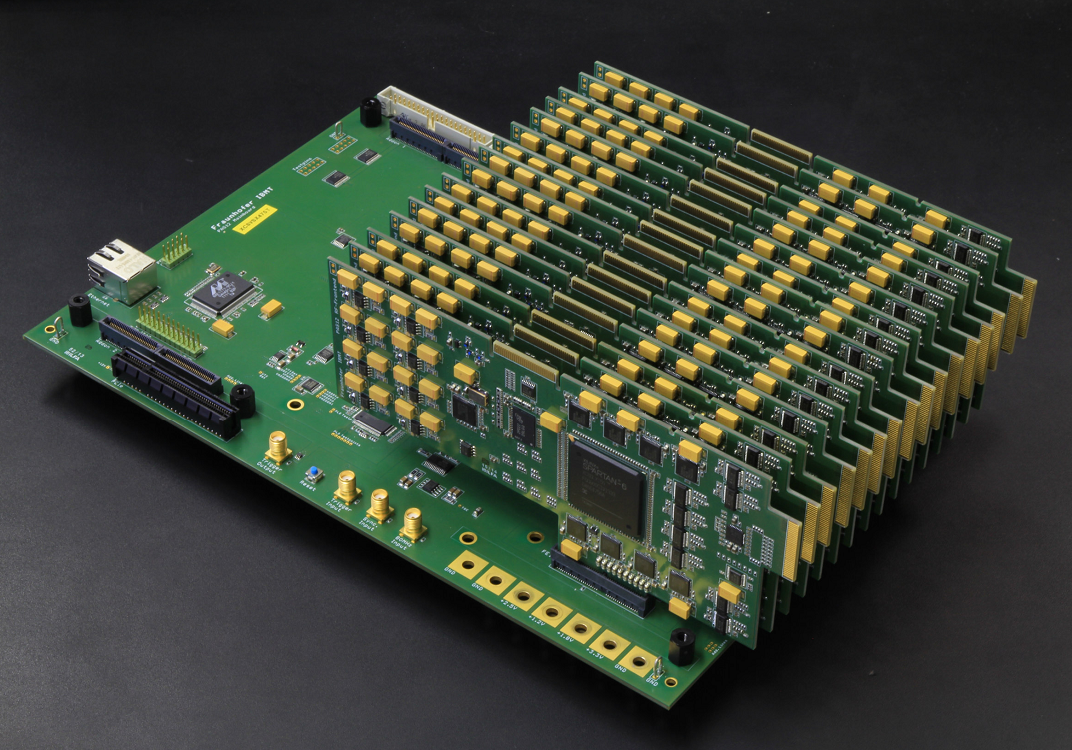
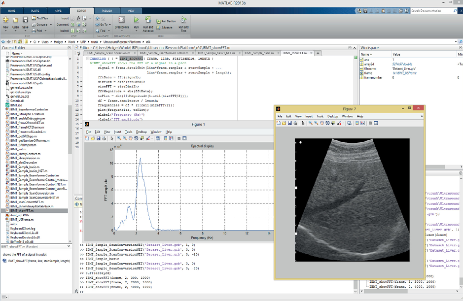
The ultrasound research system "DiPhAS", which is in its ninth generation of development, provides full control over all beamforming and imaging parameters. DiPhAS is controlled and programmed by a standard PC. As well as the basic routines provided by its operating system additional extended functions for control and processing are implemented. The system is based on a PC platform with Microsoft Windows operating system, GPU-powered for massive parallel processing (like beamforming) and includes programming manual for all interface functions with all SDK software samples. Documentation of electrical safety, system components up to layouts for OEM can be provided and a user/application specific medical certification testing is available as an option.